KC-4317

TF-1-NFκB-Luc2-DR3-Cell-Line

×
请在浏览器中启用JavaScript来完成此表单。
44359
Home » TF-1-NFκB-Luc2-DR3-Cell-Line

Background of TF-1-NFκB-Luc2-DR3-Cell-Line

TL1A and its functional receptor DR3 are members of the TNF/TNFR superfamilies of proteins.It is expressed preferentially in the tissues enriched in lymphocytes, and it may play a role in regulating lymphocyte homeostasis. This receptor has been shown to stimulate NF-kappa B activity and regulate cell apoptosis. The signal transduction of this receptor is mediated by various death domain containing adaptor proteins.NF-κB pathway consists of canonical and non-canonical pathways. The canonical NF-κB is activated by various stimuli, transducing a quick but transient transcriptional activity, to regulate the expression of various proinflammatory genes and also serve as the critical mediator for inflammatory response. Meanwhile, the activation of the non-canonical NF-κB pathway occurs through a handful of TNF receptor superfamily members.

Specifications

Catalog NumberKC-4317
Cell Line NameTF-1-NFκB-Luc2-DR3-Cell-Line
Host Cell LineTF-1-NFκB-Luc2
DescriptionStable TF-1 cell line expressing exogenous luciferase gene under the control of DR3 signaling pathway
QuantityOne vial of frozen cells (≥2-106/vial)
StabilityStable in culture over a minimum of 10 passages
ApplicationDrug screening and biological assays
Freezing MediumRPMI1640 + 20% FBS + 10% DMSO
Propagation MediumRPMI1640 + 10% FBS+5ng/mL rhGM-CSF+100μg/mL Hygromycin B+1μg/mL puromycin
Selection MarkerHygromycin B and puromycin
MorphologyMostly single, round (some polymorph) cells in suspension
SubcultureSplit saturated culture 1:4-1:8 every 2-3 days; seed out at about 1-3 × 10⁵ cells/mL
Incubation37 °C with 5% CO2
StorageLiquid nitrogen immediately upon receiving
Doubling TimeApproximately 30 hours
Mycoplasma StatusNegative
In Vivo ValidationNA

Cell Line Generation

TF-1-NFκB-Luc2-DR3 cell line was generated using lentivirus expressing DR3 sequence.

Characterization

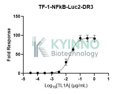
Figure 1. TF-1-NFκB-Luc2-DR3 cell line was seeded into the 96-well plate, and treated with TL1A at a maximum concentration of 1μg/mL for 6 hours, then readout with Bright-Glo system.

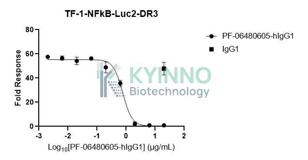
Figure 2. TF-1-NFκB-Luc2-DR3 cell line was seeded into the 96-well plate, and treated with PF-06480605-hIgG1 at a maximum concentration 20μg/mL for 1 hours, then treated with TL1A (100ng/mL), incubated for 6 hours and readout with Bright-Glo system.

Cell Resuscitation

  1. Prewarm culture medium (RPMI1640 supplemented with 10% FBS, 5ng/mL rhGM-CSF, 100μg/mL Hygromycin and 1μg/mL Puromycin)in a 37°C water bath.
  2. Thaw the frozen vial in a 37°C water bath for 1-2 minutes.
  3. Transfer the vial into biosafety cabinet, and wipe the surface with 70% ethanol.
  4. Unscrew the top of the vial and transfer the cell suspension gently into a sterile centrifuge tube containing 9.0mL complete culture medium.
  5. Spin at ~ 125 × g for 5-7 minutes at room temperature, and discard the supernatant without disturbing the pellet.
  6. Resuspend cell pellet with the appropriate volume of complete medium and transfer the cell suspension into a T25 culture flask.
  7. Incubate the flask at 37°C, 5% CO2 incubator.
  8. Split saturated culture 1:4-1:8 every 2-3 days; seed out at about 1-3 × 10⁵ cells/mL.

Cell Freezing

  1. Prepare the freezing medium (70% RPMI1640 + 20% FBS + 10% DMSO) fresh immediately before use.
  2. Keep the freezing medium on ice and label cryovials.
  3. Transfer cells to a sterile, conical centrifuge tube, and count the cells.
  4. Centrifuge the cells at 250×g for 5 minutes at room temperature and carefully aspirate off the medium.
  5. Resuspend the cells at a density of at least 3×10⁶ cells/mL in chilled freezing medium.
  6. Aliquot 1 mL of the cell suspension into each cryovial.
  7. Freeze cells in the CoolCell freezing container overnight in a -80°C freezer.
  8. Transfer vials to liquid nitrogen for long-term storage.

References

  1. Valatas V, Kolios G, Bamias G. TL1A (TNFSF15) and DR3 (TNFRSF25): A Co-stimulatory System of Cytokines With Diverse Functions in Gut Mucosal Immunity. Front Immunol. 2019 Mar 27;10:583. doi: 10.3389/fimmu.2019.00583. PMID: 30972074; PMCID: PMC6445966.
  2. Chinnaiyan AM, O'Rourke K, Yu GL, Lyons RH, Garg M, Duan DR, Xing L, Gentz R, Ni J, Dixit VM. Signal transduction by DR3, a death domain-containing receptor related to TNFR-1 and CD95. Science. 1996 Nov 8;274(5289):990-2. doi: 10.1126/science.274.5289.990. PMID: 8875942.

Use License Agreement

Research Use Only.
Not for use in diagnostic procedures or therapeutic applications.
Redistribution of the cell line or its derivatives is prohibited without prior written permission from Kyinno Biotechnology.
请在浏览器中启用JavaScript来完成此表单。