KC-4576

Ba/F3-MPL-JAK2-V617F-GFP-Luc2-Cell-Line

×
请在浏览器中启用JavaScript来完成此表单。
53348
Home » Ba/F3-MPL-JAK2-V617F-GFP-Luc2-Cell-Line

Background of Ba/F3-MPL-JAK2-V617F-GFP-Luc2-Cell-Line

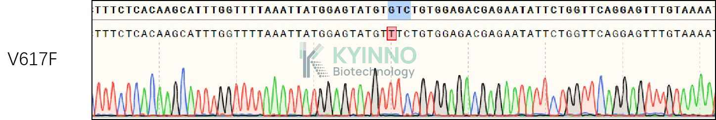
Janus kinase 2 (commonly called JAK2) is a non-receptor tyrosine kinase. It is a member of the Janus kinase family and has been implicated in signaling by members of the type II cytokine receptor family (e.g. interferon receptors), the GM-CSF receptor family (IL-3R, IL-5R and GM-CSF-R), the gp130 receptor family (e.g., IL-6R), and the single chain receptors (e.g. Epo-R, Tpo-R, GH-R, PRL-R). Mutations in JAK2 have been implicated in polycythemia vera, essential thrombocythemia, and myelofibrosis as well as other myeloproliferative disorders. This mutation (V617F), a change of valine to phenylalanine at the 617 position, appears to render hematopoietic cells more sensitive to growth factors such as erythropoietin and thrombopoietin, because the receptors for these growth factors require JAK2 for signal transduction. An inhibitor of JAK2-STAT5, AZD1480, was pointed as having activity in primary and CRPC. Jak2 mutation, when demonstrable, is one of the methods of diagnosing polycythemia vera.
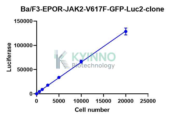
Ba/F3 cell, a murine interleukin-3 dependent pro-B cell line, is a popular system for exploring both kinases and their inhibitors, because some protein kinases can render the Ba/F3 cells to be depended on the activation of the kinases instead of IL-3 supplement, while their inhibitors can antagonize the kinase-dependent growth effects.
Luciferase is an oxidative enzyme that can produce bioluminescence with addition of luciferin, but doesn’t need an external light source unlike fluorescent proteins. Photo emission can be detected directly by light sensitive devices. Such as luminometers or modified microscopes. Luciferase is widely used in many fields of biological research, such as transcriptional activity, kinase or other enzyme activity, cellular ATP level, and whole animal imaging.

Specifications

Catalog NumberKC-4576
Cell Line NameBa/F3-MPL-JAK2-V617F-GFP-Luc2-Cell-Line
Clone Number4#
Host Cell LineBa/F3
DescriptionStable Ba/F3 clone expressing exogenous MPL-JAK2 bearing V617F-GFP-Luc2 amino acid mutation
QuantityOne vial of frozen cells (≥2-106/vial)
StabilityStable in culture over a minimum of 10 passages
ApplicationDrug screening and biological assays
Freezing Medium70% RPMI1640+20% FBS+10% DMSO
Propagation MediumRPMI1640+10%FBS
Selection MarkerNA
MorphologyMostly single, round (some polymorph) cells in suspension
SubcultureSplit saturated culture 1:10 every 3 days; seed out at about 1-3 × 10⁵ cells/mL
Incubation37 °C with 5% CO2
StorageLiquid nitrogen immediately upon receiving
Doubling TimeApproximately 20 hours
Mycoplasma StatusNegative

Cell Line Generation

Ba/F3-MPL-JAK2-V617F-GFP-Luc2-cell-Line was generated using retrovirus vector expressing human MPL-JAK2-V617F-GFP-Luc2 sequence.

Characterization

Figure 1: Characterization of V617F in the Ba/F3-MPL-JAK2-V617F-GFP-Luc2 stable clone using PCR sequencing.

Figure 2: Figure 1: Characterization of the Ba/F3-MPL-JAK2-V617F-GFP-Luc2-cell-line stable clone using Bright-Lite Luciferase Assay System in the conditions of different cell number.

Cell Resuscitation

1. Prewarm culture medium (RPMI1640 + 10% FBS)in a 37°C water bath.
2. Thaw the frozen vial in a 37°C water bath for 1-2 minutes.
3. Transfer the vial into biosafety cabinet, and wipe the surface with 70% ethanol.
4. Unscrew the top of the vial and transfer the cell suspension gently into a sterile centrifuge tube containing 9.0mL complete culture medium.
5. Spin at ~ 125 × g for 5-7 minutes at room temperature, and discard the supernatant without disturbing the pellet.
6. Resuspend cell pellet with the appropriate volume of complete medium and transfer the cell suspension into a T25 culture flask.
7. Incubate the flask at 37°C, 5% CO2 incubator.
8. Split saturated culture 1:10 every 3 days; seed out at about 1-3 × 10⁵ cells/mL.

Cell Freezing

1. Prepare the freezing medium (70% RPMI-1640 + 20% FBS + 10% DMSO) fresh immediately before use.
2. Keep the freezing medium on ice and label cryovials.
3. Transfer cells to a sterile, conical centrifuge tube, and count the cells.
4. Centrifuge the cells at 250×g for 5 minutes at room temperature and carefully aspirate off the medium.
5. Resuspend the cells at a density of at least 3×10⁶ cells/mL in chilled freezing medium.
6. Aliquot 1 mL of the cell suspension into each cryovial.
7. Freeze cells in the CoolCell freezing container overnight in a -80°C freezer.
8. Transfer vials to liquid nitrogen for long-term storage.

References

1. Bole-Feysot C, Goffin V, Edery M, Binart N, Kelly PA (June 1998). "Prolactin (PRL) and its receptor: actions, signal transduction pathways and phenotypes observed in PRL receptor knockout mice". Endocrine Reviews. 19 (3): 225ÿ68.
2.Brooks AJ, Dai W, O'Mara ML, Abankwa D, Chhabra Y, Pelekanos RA, et al. (2014). "Mechanism of activation of protein kinase JAK2 by the growth hormone receptor". Science. 344 (6185): 1249783.
3.Kralovics R, Passamonti F, Buser AS, Teo SS, Tiedt R, Passweg JR, Tichelli A, Cazzola M, Skoda RC (April 2005). "A gain-of-function mutation of JAK2 in myeloproliferative disorders". The New England Journal of Medicine. 352 (17): 1779ÿ90.
4.Greer LF, Szalay AA (2002). "Imaging of light emission from the expression of luciferases in living cells and organisms: a review". Luminescence. 17 (1): 43–74. doi:10.1002/bio.676. PMID 11816060.

Use License Agreement

Research Use Only.
Not for use in diagnostic procedures or therapeutic applications.
Redistribution of the cell line or its derivatives is prohibited without prior written permission from Kyinno Biotechnology.
请在浏览器中启用JavaScript来完成此表单。