KC-5101

HeLa-ERBB2-KO Cell Line

×
Please enable JavaScript in your browser to complete this form.
54190
Home » HeLa-ERBB2-KO Cell Line

Background of HeLa-ERBB2-KO Cell Line

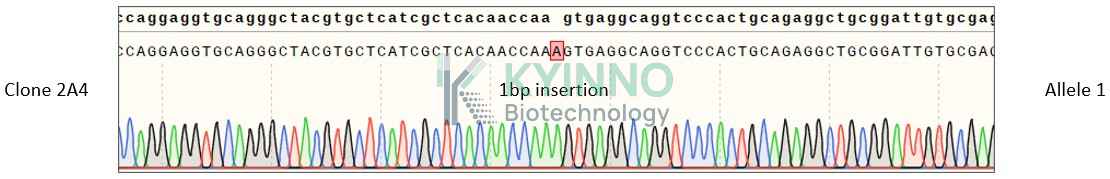
\"The ERBB2 gene, also known as HER2, encodes a tyrosine kinase receptor belonging to the epidermal growth factor receptor (EGFR) family. This protein has no ligand binding domain of its own and therefore cannot bind growth factors. However, it does bind tightly to other ligand-bound EGF receptor family members to form a heterodimer, stabilizing ligand binding and enhancing kinase-mediated activation of downstream signalling pathways, such as those involving mitogen-activated protein kinase and phosphatidylinositol-3 kinase. Amplification and/or overexpression of this gene has been reported in numerous cancers, including breast and ovarian tumors. Alternative splicing results in several additional transcript variants, some encoding different isoforms and others that have not been fully characterized. \"

Specifications

Catalog NumberKC-5101
Cell Line NameHeLa-ERBB2-KO Cell Line
Clone Number2A4
Host Cell LineHeLa
DescriptionStable HeLa clone with human ERBB2 gene knockout, No.2A4
QuantityTwo vials of frozen cells (≥2-106/vial)
StabilityStable in culture over a minimum of 10 passages
ApplicationDrug screening and biological assays
Freezing MediumDMEM+20% FBS+10% DMSO
Propagation MediumDMEM+10% FBS
Selection MarkerNA
Morphologyepithelial
SubcultureSplit saturated culture 1:6-1:10 every 2-3 days; seed out at about 1-3 × 10⁵ cells/mL
Incubation37 °C with 5% CO2
StorageLiquid nitrogen immediately upon receiving
Doubling TimeApproximately 30 hours
Mycoplasma StatusNegative

Cell Line Generation

HeLa-ERBB2-KO cell line was generated using the CRISPR method.

Characterization

Figure 1: Characterization of HeLa-ERBB2-KO cell line stable clone using PCR sequencing.

Figure 2: Characterization of HeLa-ERBB2-KO cell line stable clone using RT-PCR sequencing.

Figure 3: Characterization of HeLa-ERBB2-KO cell line stable clone using FACS.

Cell Resuscitation

  1. Prewarm culture medium (DMEM + 10% FBS) in a 37°C water bath.
  2. Thaw the frozen vial in a 37°C water bath for 1-2 minutes.
  3. Transfer the vial into biosafety cabinet, and wipe the surface with 70% ethanol.
  4. Unscrew the top of the vial and transfer the cell suspension gently into a sterile centrifuge tube containing 9.0mL complete culture medium.
  5. Spin at ~ 125 × g for 5-7 minutes at room temperature, and discard the supernatant without disturbing the pellet.
  6. Resuspend cell pellet with the appropriate volume of complete medium and transfer the cell suspension into a T25 culture flask.
  7. Incubate the flask at 37°C, 5% CO2 incubator.
  8. Split saturated culture 1:6-1:10 every 2-3 days; seed out at about 1-3 ×10⁵ cells/mL.

Cell Freezing

  1. Prepare the freezing medium (70% DMEM + 20% FBS + 10% DMSO) fresh immediately before use.
  2. Keep the freezing medium on ice and label cryovials.
  3. Transfer cells to a sterile, conical centrifuge tube, and count the cells.
  4. Centrifuge the cells at 250×g for 5 minutes at room temperature and carefully aspirate off the medium.
  5. Resuspend the cells at a density of at least 3×10⁶ cells/mL in chilled freezing medium.
  6. Aliquot 1 mL of the cell suspension into each cryovial.
  7. Freeze cells in the CoolCell freezing container overnight in a -80°C freezer.
  8. Transfer vials to liquid nitrogen for long-term storage.

References

  1. Pilati C, Soulabaille A, Gallois C, Blons H, Cayre A, Sroussi M, Le Corre D, Mouillet-Richard S, Mulot C, Le Malicot K, De Reynies A, Bachet JB, Borg C, Di Fiore F, Guimbaud R, Bennouna J, André T, Taieb J, Penault-Llorca F, Laurent-Puig P. ERBB2 Comprehensive Profiling and Prognostication in Stage III Colon Cancer: Findings From PETACC8 and IDEA-France Cohorts. Gastroenterology. 2025 Apr;168(4):714-724.e4. doi: 10.1053/j.gastro.2024.10.046. Epub 2024 Nov 28. PMID: 39612956.
  2. https://www.ncbi.nlm.nih.gov/gene/2064
Please enable JavaScript in your browser to complete this form.