KC-6602

293T-BRE-Luc2-Cell-Line

×
请在浏览器中启用JavaScript来完成此表单。
64216
Home » 293T-BRE-Luc2-Cell-Line

Background of 293T-BRE-Luc2-Cell-Line

Bone morphogenetic protein (BMP)/carriers approved for orthopedic procedures achieve efficacy superior or equivalent to autograft bone. It initiates the canonical BMP signaling cascade by associating with type I receptor BMPR1A and type II receptor BMPR2. Once all three components are bound together in a complex at the cell surface, BMPR2 phosphorylates and activates BMPR1A. In turn, BMPR1A propagates signal by phosphorylating SMAD1/5/8 that travel to the nucleus and act as activators and repressors of transcription of target genes. It also acts to promote expression of HAMP, via the interaction with its receptor BMPR1A/ALK3. It can also signal through non-canonical pathways such as ERK/MAP kinase signaling cascade that regulates osteoblast differentiation.

Specifications

Catalog NumberKC-6602
Cell Line Name293T-BRE-Luc2-Cell-Line
Clone Number2C1
Host Cell Line293T
DescriptionStable 293T cell line expressing exogenous luciferase gene under the control of BRE signaling pathway.
QuantityOne vial of frozen cells (≥2-106/vial)
StabilityStable in culture over a minimum of 10 passages
ApplicationDrug screening and biological assays
Freezing Medium70% DMEM+20% FBS+10% DMSO
Propagation MediumDMEM+10% FBS+150µg/mL Hygromycin B
Selection MarkerHygromycin B
MorphologyEpithelial
SubcultureSplit saturated culture 1:4-1:5 every 2-3 days; seed out at about 1-3 × 10⁵ cells/mL
Incubation37 °C with 5% CO2
StorageLiquid nitrogen immediately upon receiving
Doubling TimeApproximately 30 hours
Mycoplasma StatusNegative

Cell Line Generation

293T-BRE-Luc2 Cell Line was generated using a lentiviral vector expressing BRE sequence.

Characterization

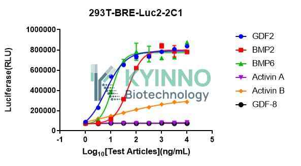
Figure 1. 293T-BRE-Luc2 cell line was seed into the 96-well plate, and treated with different ligands at a maximum concentration 10000 ng/mL diluted 3.16-fold for 16 hours, then readout with Bright-Glo system.

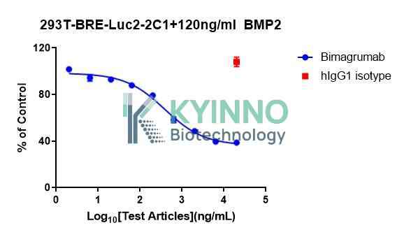
Figure 2. 293T-BRE-Luc2 cell line was seed into the 96-well plate, and treated with Bimagrumab at a maximum concentration 20 μg/mL diluted 3.16-fold for 1 hours, and then BMP2 treatment in the concentrations of 120 ng/mL for 16 h, then readout with Bright-Glo system.

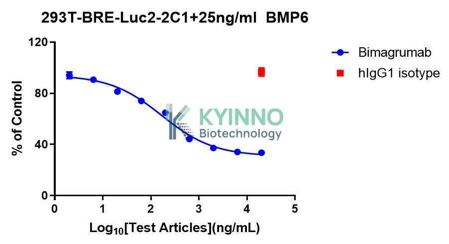
Figure 3. 293T-BRE-Luc2 cell line was seed into the 96-well plate, and treated with Bimagrumab at a maximum concentration 20 μg/mL diluted 3.16-fold for 1 hours, and then BMP6 treatment in the concentrations of 25 ng/mL for 16 h, then readout with Bright-Glo system.

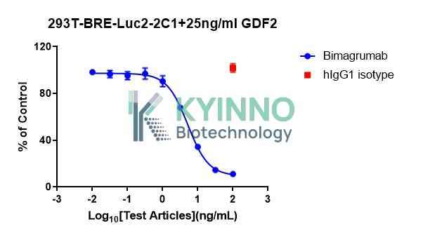
Figure 4. 293T-BRE-Luc2 cell line was seed into the 96-well plate, and treated with Bimagrumab at a maximum concentration 20 μg/mL diluted 3.16-fold for 1 hours, and then GDF2 treatment in the concentrations of 25 ng/mL for 16 h, then readout with Bright-Glo system.

Cell Resuscitation

1. Prewarm culture medium (DMEM supplemented with 10% FBS, 150µg/mL Hygromycin B)in a 37°C water bath.
2. Thaw the frozen vial in a 37°C water bath for 1-2 minutes.
3. Transfer the vial into biosafety cabinet, and wipe the surface with 70% ethanol.
4. Unscrew the top of the vial and transfer the cell suspension gently into a sterile centrifuge tube containing 9.0mL complete culture medium.
5. Spin at ~ 125 × g for 5-7 minutes at room temperature, and discard the supernatant without disturbing the pellet.
6. Resuspend cell pellet with the appropriate volume of complete medium and transfer the cell suspension into a T25 culture flask.
7. Incubate the flask at 37°C, 5% CO2 incubator.
8. Split saturated culture 1:4-1:5 every 2-3 days; seed out at about 1-3 × 10⁵ cells/mL.

Cell Freezing

1. Prepare the freezing medium (70% DMEM + 20% FBS + 10% DMSO) fresh immediately before use.
2. Keep the freezing medium on ice and label cryovials.
3. Transfer cells to a sterile, conical centrifuge tube, and count the cells.
4. Centrifuge the cells at 250×g for 5 minutes at room temperature and carefully aspirate off the medium.
5. Resuspend the cells at a density of at least 3×10⁶ cells/mL in chilled freezing medium.
6. Aliquot 1 mL of the cell suspension into each cryovial.
7. Freeze cells in the CoolCell freezing container overnight in a -80°C freezer.
8. Transfer vials to liquid nitrogen for long-term storage.

References

1.Seeherman HJ, Berasi SP, Brown CT, Martinez RX, Juo ZS, Jelinsky S, Cain MJ, Grode J, Tumelty KE, Bohner M, Grinberg O, Orr N, Shoseyov O, Eyckmans J, Chen C, Morales PR, Wilson CG, Vanderploeg EJ, Wozney JM. A BMP/activin A chimera is superior to native BMPs and induces bone repair in nonhuman primates when delivered in a composite matrix. Sci Transl Med. 2019 Apr 24;11(489):eaar4953. doi: 10.1126/scitranslmed.aar4953. PMID: 31019025.
2. Keller S, Nickel J, Zhang JL, Sebald W, Mueller TD. Molecular recognition of BMP-2 and BMP receptor IA. Nat Struct Mol Biol. 2004 May;11(5):481-8. doi: 10.1038/nsmb756. Epub 2004 Apr 4. PMID: 15064755.
3. Wang CY, Xu Y, Traeger L, Dogan DY, Xiao X, Steinbicker AU, Babitt JL. Erythroferrone lowers hepcidin by sequestering BMP2/6 heterodimer from binding to the BMP type I receptor ALK3. Blood. 2020 Feb 6;135(6):453-456. doi: 10.1182/blood.2019002620. PMID: 31800957; PMCID: PMC7005366.

Use License Agreement

Research Use Only.
Not for use in diagnostic procedures or therapeutic applications.
Redistribution of the cell line or its derivatives is prohibited without prior written permission from Kyinno Biotechnology.
请在浏览器中启用JavaScript来完成此表单。