KC-6735

KMS12-BM-GFP-Luc2 Cell Line

×
请在浏览器中启用JavaScript来完成此表单。
65478
Home » KMS12-BM-GFP-Luc2 Cell Line

Background of KMS12-BM-GFP-Luc2 Cell Line

Luciferase is an oxidative enzyme that can produce bioluminescence with addition of luciferin, but doesn’t need an external light source unlike fluorescent proteins. Photo emission can be detected directly by light sensitive devices. Such as luminometers or modified microscopes. Luciferase is widely used in many fields of biological research, such as transcriptional activity, kinase or other enzyme activity, cellular ATP level, and whole animal imaging.

Specifications

Catalog NumberKC-6735
Cell Line NameKMS12-BM-GFP-Luc2 Cell Line
NCBI/UniProt Accession NumberNA
Clone Number1#
Host Cell LineKMS12-BM
DescriptionKMS12-BM cell line stably expressing exogenous GFP-Luc2 gene
QuantityTwo vials of frozen cells (≥2-106/vial)
StabilityStable in culture over a minimum of 10 passages
ApplicationDrug screening and biological assays
Freezing Medium70% RPMI1640 + 20% FBS + 10% DMSO
Propagation MediumRPMI1640 + 10% FBS
Selection MarkerNA
MorphologySuspension
SubcultureSplit saturated culture 1:2-1:4 every 2-3 days; seed out at about 1-2 × 10⁵ cells/mL
Incubation37 °C with 5% CO2
StorageLiquid nitrogen immediately upon receiving
Doubling TimeApproximately 60 hours
Mycoplasma StatusNegative

Cell Line Generation

KMS12-BM-GFP-Luc2 cell line was generated using a lentiviral vector expressing the GFP-Luc2 sequence.

Characterization

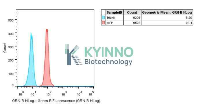
Figure 1: Characterization of GFP overexpression in KMS12-BM-GFP-Luc2 stable clones using FACS.

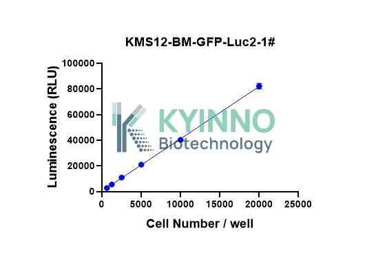
Figure 2: Characterization of the KMS12-BM-GFP-Luc2 Cell Line stable clone using Bright-Lite Luciferase Assay System in the conditions of different cell number.

Cell Resuscitation

  1. Prewarm culture medium (RPMI1640 supplemented with 10% FBS) in a 37°C water bath.
  2. Thaw the frozen vial in a 37°C water bath for 1-2 minutes.
  3. Transfer the vial into biosafety cabinet, and wipe the surface with 70% ethanol.
  4. Unscrew the top of the vial and transfer the cell suspension gently into a sterile centrifuge tube containing 9.0 mL complete culture medium.
  5. Spin at ~ 125 × g for 5-7 minutes at room temperature, and discard the supernatant without disturbing the pellet.
  6. Resuspend cell pellet with the appropriate volume of complete medium and transfer the cell suspension into a T25 culture flask.
  7. Incubate the flask at 37°C, 5% CO2 incubator.
  8. Split saturated culture 1:2-1:4 every 2-3 days; seed out at about 1-2 × 10⁵ cells/mL.

Cell Freezing

  1. Prepare the freezing medium (70% RPMI1640 + 20% FBS + 10% DMSO) fresh immediately before use.
  2. Keep the freezing medium on ice and label cryovials.
  3. Transfer cells to a sterile, conical centrifuge tube, and count the cells.
  4. Centrifuge the cells at 250×g for 5 minutes at room temperature and carefully aspirate off the medium.
  5. Resuspend the cells at a density of at least 3×10⁶ cells/mL in chilled freezing medium.
  6. Aliquot 1 mL of the cell suspension into each cryovial.
  7. Freeze cells in the CoolCell freezing container overnight in a -80°C freezer.
  8. Transfer vials to liquid nitrogen for long-term storage.

References

  1. Kim SB, Fujii R. A New Lineage of Artificial Luciferases for Mammalian Cell Imaging. Methods Mol Biol. 2021;2274:43-51. doi: 10.1007/978-1-0716-1258-3_5. PMID: 34050461.

Use License Agreement

Research Use Only.
Not for use in diagnostic procedures or therapeutic applications.
Redistribution of the cell line or its derivatives is prohibited without prior written permission from Kyinno Biotechnology.
请在浏览器中启用JavaScript来完成此表单。