KC-2991

MKN45-CLDN18.2-PDL1 Cell Line

×
Please enable JavaScript in your browser to complete this form.
26851
Home » MKN45-CLDN18.2-PDL1 Cell Line

Background of MKN45-CLDN18.2-PDL1 Cell Line

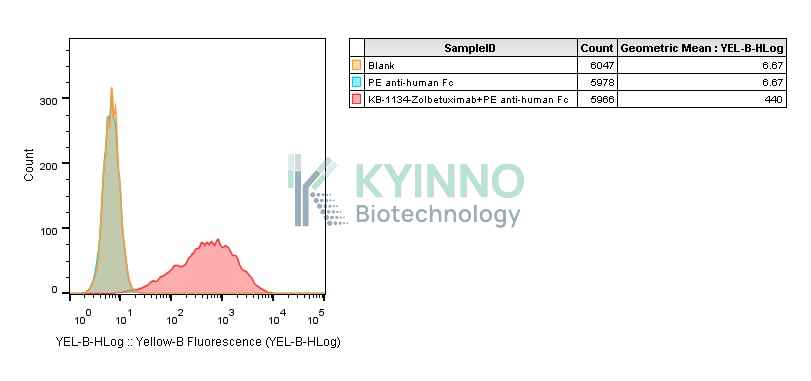
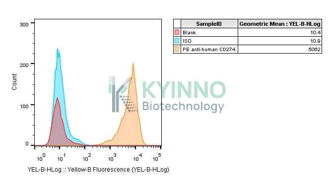
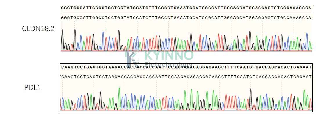
The claudin18.2 (CLDN18.2) protein, an isoform of claudin18, a member of the tight junction protein family, is a highly selective biomarker with limited expression in normal tissues and often abnormal expression during the occurrence and development of various primary malignant tumors, such as gastric cancer/gastroesophageal junction (GC/GEJ) cancer, breast cancer, colon cancer, liver cancer, head and neck cancer, bronchial cancer and non-small-cell lung cancer. CLDN18.2 participates in the proliferation, differentiation and migration of tumor cells. Recent studies have identified CLDN18.2 expression as a potential specific marker for the diagnosis and treatment of these tumors. Zolbetuximab (claudiximab, IMAB362), a monoclonal antibody (mAb) against CLDN18.2, have been developed. The engagement of programmed cell death protein 1 (PD-1; encoded by the PDCD1 gene) receptor expressed on activated T cells and its ligand programmed death-ligand 1 (PD-L1; encoded by the CD274 gene) is a major co-inhibitory checkpoint signaling that controls T-cell activities. Various types of cancers express high levels of PD-L1 and exploit the PD-L1/PD-1 signaling to evade T-cell immunity. Blocking the PD-L1/PD-1 pathway has consistently shown remarkable anti-tumor effects in patients with advanced cancers and is recognized as the gold standard for developing new immune checkpoint blockade (ICB) and combination therapies.

Specifications

Catalog NumberKC-2991
Cell Line NameMKN45-CLDN18.2-PDL1 Cell Line
Clone Number6#
Host Cell LineMKN45
DescriptionStable MKN45 cell line expressing exogenous human CLDN18.2 and PDL1 gene
QuantityTwo vials of frozen cells (≥2-106/vial)
StabilityStable in culture over a minimum of 10 passages
ApplicationDrug screening and biological assays
Freezing Medium70% RPMI1640 + 20% FBS + 10% DMSO
Propagation MediumRPMI1640 + 10% FBS + 1μg/mL Puromycin+300μg/mL Hygromycin B
Selection MarkerPuromycin | Hygromycin B
MorphologyFibroblastoid cells growing as a monolayer
SubcultureSplit saturated culture 1:3-1:4 every 2-3 days; seed out at about 1-3 × 10⁵ cells/mL
Incubation37 °C with 5% CO2
StorageLiquid nitrogen immediately upon receiving
Doubling TimeApproximately 30 hours
Mycoplasma StatusNegative
In Vivo ValidationNA

Cell Line Generation

MKN45-Human-CLDN18.2-PDL1 cell line was generated using a lentiviral vector expressing the human CLDN18.2 and PDL1 sequence.

Characterization

Figure 1: Characterization of CLND18.2 overexpression in MKN45 stable clones using FACS.

Figure 2: Characterization of human PDL1 overexpression in MKN45-Human-CLDN18.2-PDL1 stable clones using FACS.

Figure 3: Characterization of human CLDN18.2 and PDL1 overexpression in MKN45-Human-CLDN18.2-PDL1 stable clones using PCR Sequencing.

Cell Resuscitation

1. Prewarm culture medium (RPMI1640 + 10% FBS + 1μg/mL Puromycin+300μg/mL Hygromycin B)in a 37°C water bath. 2. Thaw the frozen vial in a 37°C water bath for 1-2 minutes. 3. Transfer the vial into biosafety cabinet, and wipe the surface with 70% ethanol. 4. Unscrew the top of the vial and transfer the cell suspension gently into a sterile centrifuge tube containing 9.0mL complete culture medium. 5. Spin at ~ 125 × g for 5-7 minutes at room temperature, and discard the supernatant without disturbing the pellet. 6. Resuspend cell pellet with the appropriate volume of complete medium and transfer the cell suspension into a T25 culture flask. 7. Incubate the flask at 37°C, 5% CO2 incubator. 8. Split saturated culture 1:3-1:4 every 2-3 days; seed out at about 1-3 × 10⁵ cells/mL.

Cell Freezing

1. Prepare the freezing medium (70% RPMI1640 + 20% FBS + 10% DMSO) fresh immediately before use. 2. Keep the freezing medium on ice and label cryovials. 3. Transfer cells to a sterile, conical centrifuge tube, and count the cells. 4. Centrifuge the cells at 250×g for 5 minutes at room temperature and carefully aspirate off the medium. 5. Resuspend the cells at a density of at least 3×10⁶ cells/mL in chilled freezing medium. 6. Aliquot 1 mL of the cell suspension into each cryovial. 7. Freeze cells in the CoolCell freezing container overnight in a -80°C freezer. 8. Transfer vials to liquid nitrogen for long-term storage.

References

1. Cao W, Xing H, Li Y, Tian W, Song Y, Jiang Z, Yu J. Claudin18.2 is a novel molecular biomarker for tumor-targeted immunotherapy. Biomark Res. 2022 May 31;10(1):38. doi: 10.1186/s40364-022-00385-1. PMID: 35642043; PMCID: PMC9153115. 2. Cha JH, Chan LC, Li CW, Hsu JL, Hung MC. Mechanisms Controlling PD-L1 Expression in Cancer. Mol Cell. 2019 Nov 7;76(3):359-370. doi: 10.1016/j.molcel.2019.09.030. Epub 2019 Oct 24. PMID: 31668929; PMCID: PMC6981282.
Please enable JavaScript in your browser to complete this form.