KC-1478

NALM6-Luc2 Cell Line

×
Please enable JavaScript in your browser to complete this form.
22217
Home » NALM6-Luc2 Cell Line

Background of NALM6-Luc2 Cell Line

Luciferase is an oxidative enzyme that can produce bioluminescence with addition of luciferin, but doesn’t need an external light source unlike fluorescent proteins. Photo emission can be detected directly by light sensitive devices. Such as luminometers or modified microscopes. Luciferase is widely used in many fields of biological research, such as transcriptional activity, kinase or other enzyme activity, cellular ATP level, and whole animal imaging.

Specifications

Catalog NumberKC-1478
Cell Line NameNALM6-Luc2 Cell Line
NCBI/UniProt Accession NumberN/A
Clone Numberpool
Host Cell LineNALM6
DescriptionStable NALM6-Luc2 cell line expressing exogenous luciferase gene
QuantityTwo vials of frozen cells (≥2-106/vial)
StabilityStable in culture over a minimum of 10 passages
ApplicationDrug screening and biological assays
Freezing Medium70% IMDM+20%FBS+10%DMSO
Propagation MediumIMDM+10%FBS+500µg/mL Hygromycin B
Selection MarkerHygromycin B
Morphologylymphocyte-like
SubcultureSplit saturated culture 1:3-1:6 every 2-3 days; seed out at about 1-3 × 10⁵ cells/mL
Incubation37 °C with 5% CO2
StorageLiquid nitrogen immediately upon receiving
Doubling TimeApproximately 30 hours
Mycoplasma StatusNegative
In Vivo ValidationYes

Cell Line Generation

NALM6-Luc2 Cell Line was generated using a lentiviral vector expressing luciferase sequence.

Characterization

Figure 1: Characterization of the NALM6 Luc2 cell line stable clone using Bright-Glo™ Luciferase Assay System in the conditions of different cell number.

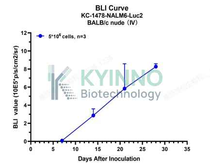
Figure 2: Bioluminescence imaging (BLI) was used to track the progression of NALM6-Luc2 tumors following intravenous (IV) inoculation of 5×10⁶ cells. Data are presented as mean ± SD (n=3).

Cell Resuscitation

  1. Prewarm culture medium (IMDM+10%FBS+500µg/mL Hygromycin B)in a 37°C water bath.
  2. Thaw the frozen vial in a 37°C water bath for 1-2 minutes.
  3. Transfer the vial into biosafety cabinet, and wipe the surface with 70% ethanol.
  4. Unscrew the top of the vial and transfer the cell suspension gently into a sterile centrifuge tube containing 9.0mL complete culture medium.
  5. Spin at ~ 125 × g for 5-7 minutes at room temperature, and discard the supernatant without disturbing the pellet.
  6. Resuspend cell pellet with the appropriate volume of complete medium and transfer the cell suspension into a T25 culture flask.
  7. Incubate the flask at 37°C, 5% CO2 incubator.
  8. Split saturated culture 1:3-1:6 every 2-3 days; seed out at about 1-3 × 10⁵ cells/mL.

Cell Freezing

  1. Prepare the freezing medium (70% IMDM+20%FBS+10%DMSO) fresh immediately before use.
  2. Keep the freezing medium on ice and label cryovials.
  3. Transfer cells to a sterile, conical centrifuge tube, and count the cells.
  4. Centrifuge the cells at 250×g for 5 minutes at room temperature and carefully aspirate off the medium.
  5. Resuspend the cells at a density of at least 3×10⁶ cells/mL in chilled freezing medium.
  6. Aliquot 1 mL of the cell suspension into each cryovial.
  7. Freeze cells in the CoolCell freezing container overnight in a -80°C freezer.
  8. Transfer vials to liquid nitrogen for long-term storage.

References

  1. Greer LF, Szalay AA (2002). Imaging of light emission from the expression of luciferases in living cells and organisms: a review. Luminescence. 17 (1): 43–74. doi:10.1002/bio.676. PMID 11816060.
Please enable JavaScript in your browser to complete this form.