KC-3905

Raji-EGFR-High-Cell-Line

×
Please enable JavaScript in your browser to complete this form.
35006
Home » Raji-EGFR-High-Cell-Line

Background of Raji-EGFR-High-Cell-Line

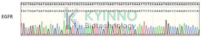
EGFR is a transmembrane glycoprotein that is one of four members of the ErbB family of tyrosine kinase receptors. Activation of EGFR leads to autophosphorylation of the receptor tyrosine kinase, which initiates downstream signaling cascades involved in the regulation of cell proliferation, differentiation, and survival. The abnormal activation of EGFR through a variety of mechanisms, such as receptor overexpression, mutation, ligand-dependent receptor dimerization, ligand-independent activation, is associated with the occurrence and development of a variety of human tumors. Inhibition of EGFR is one of the key targets of cancer chemotherapy.

Specifications

Catalog NumberKC-3905
Cell Line NameRaji-EGFR-High-Cell-Line
Host Cell LineRaji
DescriptionStable Raji cell line expressing exogenous human EGFR gene
QuantityTwo vials of frozen cells (≥2-106/vial)
StabilityStable in culture over a minimum of 10 passages
ApplicationDrug screening and biological assays
Freezing Medium70% RPMI1640+20% FBS+10% DMSO
Propagation MediumRPMI1640+10%FBS + 400 µg/mL Hygromycin B
Selection MarkerHygromycin B
MorphologyMostly single, round (some polymorph) cells in suspension
SubcultureSplit saturated culture 1:3 every 3 days; seed out at about 1-3 × 10⁵ cells/mL
Incubation37 °C with 5% CO2
StorageLiquid nitrogen immediately upon receiving
Doubling TimeApproximately 24-36 hours
Mycoplasma StatusNegative
In Vivo ValidationNA

Cell Line Generation

Raji human EGFR High cell line was generated using a lentiviral vector expressing the human EGFR sequence.

Characterization

Figure1: Characterization of EGFR overexpression in Raji stable clones using FACS.

Figure2: Characterization of EGFR and its mutants overexpressing in Raji stable clones using PCR sequencing

Cell Resuscitation

  1. Prewarm culture medium (RPMI1640 + 10% FBS + 400 µg/mL Hygromycin B)in a 37°C water bath.
  2. Thaw the frozen vial in a 37°C water bath for 1-2 minutes.
  3. Transfer the vial into biosafety cabinet, and wipe the surface with 70% ethanol.
  4. Unscrew the top of the vial and transfer the cell suspension gently into a sterile centrifuge tube containing 9.0mL complete culture medium.
  5. Spin at ~ 125 × g for 5-7 minutes at room temperature, and discard the supernatant without disturbing the pellet.
  6. Resuspend cell pellet with the appropriate volume of complete medium and transfer the cell suspension into a T25 culture flask.
  7. Incubate the flask at 37°C, 5% CO2 incubator.
  8. Split saturated culture 1:2 every 2-3 days; seed out at about 1-3 × 10⁵ cells/mL.

Cell Freezing

  1. Prepare the freezing medium (70% RPMI-1640 + 20% FBS + 10% DMSO) fresh immediately before use.
  2. Keep the freezing medium on ice and label cryovials.
  3. Transfer cells to a sterile, conical centrifuge tube, and count the cells.
  4. Centrifuge the cells at 250×g for 5 minutes at room temperature and carefully aspirate off the medium.
  5. Resuspend the cells at a density of at least 3×10⁶ cells/mL in chilled freezing medium.
  6. Aliquot 1 mL of the cell suspension into each cryovial.
  7. Freeze cells in the CoolCell freezing container overnight in a -80°C freezer.
  8. Transfer vials to liquid nitrogen for long-term storage

References

  1. ingh D, Attri BK, Gill RK, Bariwal J. Review on EGFR Inhibitors: Critical Updates. Mini Rev Med Chem. 2016;16(14):1134-66. doi: 10.2174/1389557516666160321114917. PMID: 26996617.
  2. Yarden Y, Shilo BZ. SnapShot: EGFR signaling pathway. Cell. 2007 Nov 30;131(5):1018. doi: 10.1016/j.cell.2007.11.013. PMID: 18045542.
Please enable JavaScript in your browser to complete this form.