| Catalog Number | KC-3847 |
|---|
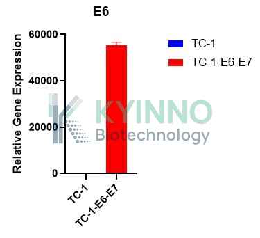
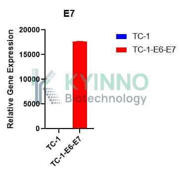
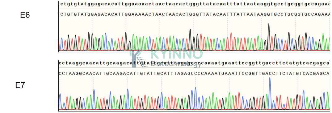
| Cell Line Name | TC1-E6-E7 Cell Line |
|---|
| Clone Number | 4# |
|---|
| Host Cell Line | TC1 |
|---|
| Description | Stable TC1 cell line expressing exogenous E6-E7 gene |
|---|
| Quantity | One vial of frozen cells (≥2-106/vial) |
|---|
| Stability | Stable in culture over a minimum of 10 passages |
|---|
| Application | Drug screening and biological assays |
|---|
| Freezing Medium | 70% RPMI1640 + 20% FBS + 10% DMSO |
|---|
| Propagation Medium | RPMI1640 + 10% FBS + 8μg/ml Puromycin |
|---|
| Selection Marker | Puromycin |
|---|
| Morphology | Epithelial |
|---|
| Subculture | Split saturated culture 1:4-1:8 every 2-3 days; seed out at about 1-3 × 10⁵ cells/mL |
|---|
| Incubation | 37 °C with 5% CO2 |
|---|
| Storage | Liquid nitrogen immediately upon receiving |
|---|
| Doubling Time | Approximately 30 hours |
|---|
| Mycoplasma Status | Negative |
|---|
| In Vivo Validation | NA |
|---|