KC-5046

U87MG-MSH2-KO Cell Line

×
Please enable JavaScript in your browser to complete this form.
54038
Home » U87MG-MSH2-KO Cell Line

Background of U87MG-MSH2-KO Cell Line

The MSH2 (MutS homolog 2) gene plays a critical role in DNA mismatch repair (MMR), a system that corrects errors occurring during DNA replication. It encodes a protein that forms heterodimers with MSH6 or MSH3 to recognize and repair base mismatches and insertion-deletion loops. Defects in MSH2 impair MMR function, leading to microsatellite instability (MSI) and increased mutation rates, which are hallmarks of Lynch syndrome (hereditary nonpolyposis colorectal cancer, HNPCC). Patients with MSH2 mutations have a high lifetime risk of colorectal, endometrial, ovarian, and other cancers.

Specifications

Catalog NumberKC-5046
Cell Line NameU87MG-MSH2-KO Cell Line
Clone Number6B1
Host Cell LineU87MG
DescriptionStable U87MG cell line with MSH2 gene knockout, No.6B1
QuantityTwo vials of frozen cells (≥2-106/vial)
StabilityStable in culture over a minimum of 10 passages
ApplicationDrug screening and biological assays
Freezing Medium70% MEM+20% FBS+10% DMSO
Propagation MediumMEM+10% FBS+0.01mM NEAA+1% Sodium Pyruvate
Selection MarkerNA
MorphologyEpithelial
SubcultureSplit saturated culture 1:4-1:6 every 2-3 days; seed out at about 1-3 × 10⁵ cells/mL
Incubation37 °C with 5% CO2
StorageLiquid nitrogen immediately upon receiving
Doubling TimeApproximately 39 hours
Mycoplasma StatusNegative

Cell Line Generation

U87MG-MSH2-KO cell line was generated using the CRISPR method.

Characterization

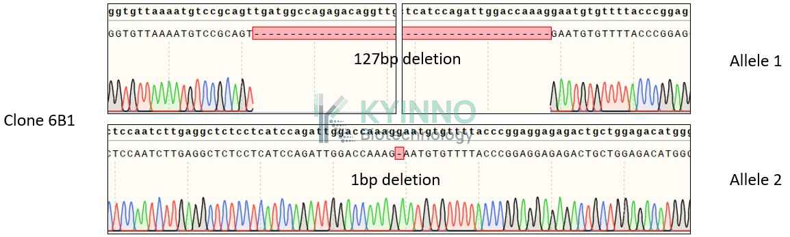
Figure 1: Characterization of U87MG-MSH2-KO Cell Line stable clone using PCR sequencing.

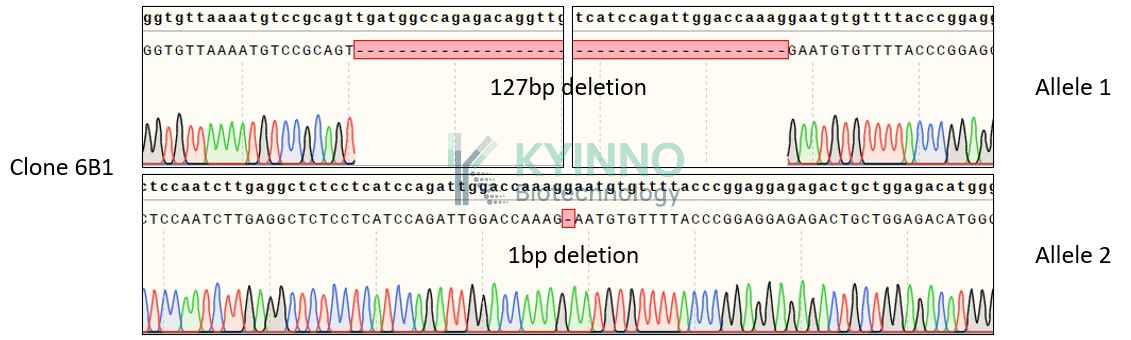
Figure 2: Characterization of U87MG-MSH2-KO Cell Line stable clone using RT-PCR sequencing.

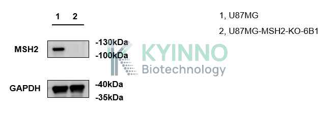
Figure 3: Characterization of U87MG-MSH2-KO Cell Line stable clone using Western blot.

Cell Resuscitation

  1. Prewarm culture medium (MEM+10% FBS+0.01mM NEAA+1% Sodium Pyruvate) in a 37°C water bath.
  2. Thaw the frozen vial in a 37°C water bath for 1-2 minutes.
  3. Transfer the vial into biosafety cabinet, and wipe the surface with 70% ethanol.
  4. Unscrew the top of the vial and transfer the cell suspension gently into a sterile centrifuge tube containing 9.0mL complete culture medium.
  5. Spin at ~ 125 × g for 5-7 minutes at room temperature, and discard the supernatant without disturbing the pellet.
  6. Resuspend cell pellet with the appropriate volume of complete medium and transfer the cell suspension into a T25 culture flask.
  7. Incubate the flask at 37°C, 5% CO2 incubator.
  8. Split saturated culture 1:4-1:6 every 2-3 days; seed out at about 1-3 × 10⁵ cells/mL.

Cell Freezing

  1. Prepare the freezing medium (70% MEM + 20% FBS + 10% DMSO) fresh immediately before use.
  2. Keep the freezing medium on ice and label cryovials.
  3. Transfer cells to a sterile, conical centrifuge tube, and count the cells.
  4. Centrifuge the cells at 250×g for 5 minutes at room temperature and carefully aspirate off the medium.
  5. Resuspend the cells at a density of at least 3×10⁶ cells/mL in chilled freezing medium.
  6. Aliquot 1 mL of the cell suspension into each cryovial.
  7. Freeze cells in the CoolCell freezing container overnight in a -80°C freezer.
  8. Transfer vials to liquid nitrogen for long-term storage.

References

  1. Boland CR, Goel A. Microsatellite instability in colorectal cancer. Gastroenterology. 2010 Jun;138(6):2073-2087.e3. doi: 10.1053/j.gastro.2009.12.064. PMID: 20420947; PMCID: PMC3037515.
  2. Lynch HT, Lynch PM, Lanspa SJ, Snyder CL, Lynch JF, Boland CR. Review of the Lynch syndrome: history, molecular genetics, screening, differential diagnosis, and medicolegal ramifications. Clin Genet. 2009 Jul;76(1):1-18. doi: 10.1111/j.1399-0004.2009.01230.x. PMID: 19659756; PMCID: PMC2846640.
Please enable JavaScript in your browser to complete this form.